Friday, 7 December 2018

AVASCULAR NECROSIS

Avascular Necrosis also called osteonecrosis, aseptic necrosis, or ischemic bone necrosis, is a condition that occurs when there is loss of blood to the bone. Since bone is the living tissue that requires blood, an interruption to the blood supply causes the bone to die. This condition is most common in people between the ages of 30 and 50.
Possible causes of avascular necrosis include:
1.      Dislocation or fracture of thighbone: This can alter the bloodstream to the bone, which can lead to shock-allied avascular necrosis. It may develop in 20% or more of people who dislocate a hip.
2.      Long use of steroid: Use of these drugs, either orally or intravenously, is associated with 35% of all cases of non-traumatic AVN. These materials gather in the blood vessels, making them narrower, and reduces the amount of blood to the bone.
3.      Extreme alcohol use: Much like corticosteroids, excessive alcohol may cause fatty substances to build in the blood vessels and decrease the blood supply to the bones.
4.      Blood clots, tenderness, and destruction to the arteries.
 Symptoms of Avascular Necrosis
In the early stages, it shows no symptoms; however, as the disease evolves it becomes painful. At first, you may get pain when you put pressure on the infected bone. Then, pain may become get constant. If the infection advances and the bone and nearby joint breakdown, you can experience severe pain that restricts the ability to use your joint.

Treatment for Avascular Necrosis

The aims of treatment for avascular necrosis is to improve the function of the infected joint, discontinue the development of bone damage, and decrease pain.
The best treatment will depend on factors, including:
  • Age
  • Stage of the disease
  • Location and amount of bone damage
  • Cause of AVN
If the cause of your avascular necrosis is found out, treatment will comprise of the efforts to manage the underlying condition. For example, if it is caused by blood clots, the doctor will recommend medicines to dissolve gobs. If swelling of the arteries is responsible, the doctor may recommend anti-inflammatory drugs.
While these nonsurgical treatments may slow the development of it, most people with the condition finally need surgery.
Medical possibilities comprises of :
  • Bone grafts, involves the removal of healthy bone from one part of the body and use it to replace the damaged bone
  • Osteotomy, a procedure that includes cutting the bone and changing the alignment to relieve stress on the bone or joint.
  • Total joint replacement, removing the damaged joint and replacing it with a synthetic joint.
  • Core decompression, a process that includes eliminating parts of the inside of the bone to release the pressure and allow new blood vessels to form.
  • Vascularized bone graft, is a technique that uses the patient's tissue to reconstruct unhealthy or broken hip joints.
Risk factors for developing avascular necrosis include:
·         Strain: Injuries, such as hip dislocation or fracture, can damage nearby blood vessels and reduce blood flow to bones.
·         Steroid: Use of high-dose corticosteroids, such as prednisone, is a common cause of avascular necrosis.
·         Excessive use of alcohol: Regular drinking for several years may create fatty deposits to form in your blood vessels.
·         Bisphosphonate use: Long period use of pills for the growth of bone density can helps to developing osteonecrosis of the jaw.
·         Some medical cures: Radiation therapy for cancer can weaken bone.
Prevention

Factors helping you prevent avascular necrosis:

·         Avoid alcohol.
·         Keep your cholesterol levels low.
·         Avoid use of high-dose steroids.

·         Do not smoke.
Wednesday, 5 December 2018

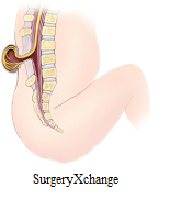
Lumbar Spine

The body part spine states to the lower back, wherever the inner spine curves towards the abdomen. "Lumbar" is ensuant from the Latin word "lumbus," that means lion, and also the body part spine earns its name. It is created for each power and adaptability.
          It has many completely different characteristics:

• The lower the bone is within the vertebral column, the additional weight it should bear. The 5 vertebrae of the body part spine (L1-L5) area unit the most important unfused vertebrae within the vertebral column, sanction native them to support the burden of the complete body part.

• The body part spine's lowest 2 spinal segments, L4-L5 and L5-S1, that embrace the vertebrae and discs, bear the foremost weight and area unit thus the foremost liable to degradation and injury.

• The body part spine meets the bone at the lumbosacral joint (L5-S1). This joint permits for significant rotation, so the pelvis and hips might swing once walking and running.

• The lower spine curves slightly inward, toward the abdomen. This inward curve of the spine is termed spinal curvature.

Lumbar bone phase

Physicians typically make a case for a patient's pathology by specializing in one bone phase, or spinal phase. The body part spine has five bone segments, termed body part phase one through five (e.g. L1, L2, L3, L4, and L5).
Each body part spine phase is comprised of:

• Two vertebrae, like L4-L5, stacked vertically with Associate in Nursing saucer between them. A healthy disc is padded, with loads of water, and contains a sponge-like substance. It acts sort of a shock within the spine, permitting flexibility and providing protection from jarring movements.

• The 2 adjacent vertebrae area unit connected within the back of the spine by 2 little joints referred to as side joints. The side joints of the body part spine permit movement to bend and twist the low back altogether directions.

• These area unit nerves that branch aloof from the vertebral column at every level of the spine. They taste little holes within the back of the lower spine. They then connect along to make the nerves, that travels into the legs down the rear of every thigh and into the calves and feet.

Doctors typically remark a patient's body part disc drawback, or nerve or alternative lower back drawback, because the level that has 2 vertebrae and also the disc between them, like L3-L4 or L4-L5. If the disc at the terribly bottom of the spine is affected, that phase is termed the lumbosacral joint L5-S1 (the S stands for sacral, that area unit the segments below the body part spine).

The following outlines the additional common causes of lower back pain:

Muscular issues

The most common reason for lower back pain is muscle strain or alternative muscle issues. Strain thanks to work, bending, or alternative arduous or repetitive use are often quite painful, however muscle strains typically heal among some days or weeks.
Degenerated Discs

Intervertebral discs area unit spongy pads that act as shock absorbers between every of the body part spine's vertebrae. Disc degeneration will produce pain within the disk space. This condition are often brought up with many various terms - on this web site it's systematically termed "degenerative disc sickness."

Lumbar Disc rupture

Herniated discs area unit commonest within the body part spine. A slipped disc might happen suddenly thanks to injury or work or happen slowly as a vicinity of general wear and tear on the spine. Leg pain (sciatica) is that the commonest symptom of a slipped disc.

Sacroiliac Joint disfunction
The sacroiliac joint, that connects with very cheap of the body part spine and also the prime of the tailbone, will cause lower back pain and/or neuralgia pain if there's any variety of disfunction within the joint that enables an excessive amount of movement or restricts traditional movement.

Spondylolisthesis

Spondylolisthesis happens once one bone slips forward over the one below it. The slip most typically happens within the lower body part vertebrae (e.g. L4 –L5 or L5 – S1). If the slipped bone compresses the nerve root at that level, it may result in leg pain and presumably foot pain.

Osteoarthritis

Osteoarthritis within the lower back is usually referred to as side joint inflammatory disease. Aging and wear and tear will cause the gristle covering the side joints within the back of the spine to become worn and worn. Excess friction will turn out bone spurs and joint swelling that cause tenderness, pressure to the nerve, and restricted vary of motion.
Lumbar stricture
The narrowing of the vertebral canal or nerve root canals (gaps wherever the nerve endings taste as they exit the spinal column) can cause the nerve endings to be squeezed, leading to leg pain, tingling, numbness, and even issue walking. This narrowing is commonly the results of bone spurs and joint swelling from degenerative joint disease.


Lumbar Spine Surgery Techniques
Lumbar decompression procedures area unit typically performed from the posterior (back) and embrace the techniques below.

Foraminotomy: once disc material and/or bone spurs press against a nerve exiting the gap (nerve passageway), a foraminotomy is also performed. Otomy is that the medical term for creating a gap. A foraminotomy makes the gap of the gap larger and relieves nervous disorder.

Laminotomy and Laminectomy: To access disc material offensive the vertebral canal, a tiny low gap is created within the plate. Through out a cutting out, all or a part of the plate is removed (ectomy is that the medical term for removal of). A cutting out is performed to alleviate funiculus compression or to achieve access to a disc from the rear. cutting out is commonly accustomed treat body part spinal stricture, a narrowing of the vertebral canal generally caused by inflammatory disease.

Facetectomy: Involves a whole or partial removal of the side joint. A facetectomy is performed to cut back nerve root compression or to achieve access to disk space.
Discectomy: Surgical removal of all or a part of a disc. Discectomy are often performed from the posterior (back) or front (anterior) through the cavity to achieve the spine.


Saturday, 1 December 2018

SPINA BIFIDA


Spina bifida through SurgeryXchange
Spina bifida is a birth defect which takes place when the spine and spinal cord don't form correctly. It comes under the wide category of neural tube defects. The neural tube is the embryonic construction that ultimately develops into the baby's brain, spinal cord, and tissues that surround them. Usually, the neural tube forms early in pregnancy, and it closes by the 28th day after formation. In babies with spina bifida, a part of the neural tube fails to grow or close properly, causes the faults in the spinal cord and in the bones of the spine.
Types of Spina Bifida
  1. Meningocele
In a form of spina bifida called meningocele, the protective tissues around the spinal cord drive out through the opening in the vertebrae, creating a sac filled with liquid. But this sac doesn't contain spinal cord, so nerve injury is less likely to happen, however, later complications are possible.
  1. Spina bifida occulta
The mildest form, spina bifida occulta results in a minor separation or gap in one or more of the bones of the spine. Several people who have it don't even know it, except the condition is discovered during an imaging test done for unrelated reasons.
  1. Myelomeningocele
It is also known as open spina bifida, it is the most severe form. The spinal canal is open along with numerous vertebrae in lower or middle back. The tissues and spinal nerves push through this opening at birth, forming a sac on the baby's back, typically exposing tissues and nerves. This makes the baby prone to life-threatening infections.
Symptoms
Following are the symptoms of spina bifida:
  1. Spina bifida occulta.Since the spinal nerves generally are not involved, typically there are no signs or symptoms. However, evident signs may sometimes be seen on the newborn's skin above the spinal defect, including an abnormal tuft of hair, or a small dimple or birthmark.
  2. The membranes near the spinal cord push out through an opening in the vertebrae, creating a sac filled with fluid, but this sac does not include the spinal cord.
  3. In this severe form of spina bifida:
    1. The spinal canal remains open along several vertebrae in the lower or middle back.
    2. Both the membranes and the spinal cord or nerves protrude at birth, forming a sac.
  • Tissues and nerves usually are exposed, though sometimes skin covers the sac.
Risk and Complications
Risks include:
  • Family history of neural tube defects. Couples who have had one child with a neural tube defect have higher chances of having another baby with the same defect. The risk increases if two previous children are having the same condition.
  • Some medications. For example, anti-seizure medications, such as valproic acid (Depakene), seem to cause neural tube defects when taken during pregnancy, possibly because they interfere with the body's ability to use folate and folic acid.
  • Women having diabetes with no control over their blood sugar level have a higher risk of having a baby with spina bifida.
  • Pre-pregnancy obesity can increase the risk of neural tube birth defects, including spina bifida.

Prevention

Get folic acid first

It is important to have a good amount of folic acid in your system during the early weeks of pregnancy to prevent spina bifida. Most women don't discover that they're pregnant until this time, experts say that all women of childbearing age should take a daily supplement of 400 micrograms (mcg) of folic acid.
Various foods, including enriched bread, pasta, rice, and some breakfast cereals, are fortified with 400 mcg of folic acid per serving.

Planning pregnancy

If you are trying to conceive, most pregnancy experts believe supplementation of at least 400 mcg of folic acid a day is the best approach for women planning a pregnancy.
Your body does not absorb folate as easily as it absorbs synthetic folic acid, and most people do not get the recommended amount of folate through diet alone, so vitamin supplements are necessary to prevent spina bifida.
To eat a healthy diet, with foods rich in folate or enriched with folic acid. This vitamin is naturally in following:
  • Citrus fruits and juices.
  • Egg yolks.
  • Dark green vegetables.

ಸೀಳು ತುಟಿಯ ಬಗ್ಗೆ ಕೇಳಿದ್ದೀರಾ?

ಸೀಳು ತುಟಿ  ಮೇಲಿನ ತುಟಿಯ ಭಾಗದಲ್ಲಿ ಸೀಳಿಕೊಂಡಿದ್ದರೆ ಅದನ್ನು ಸೀಳು ತುಟಿ ಮತ್ತು ಸೀಳು ಅಂಗುಳ  ಎಂದು ಕರೆಯಬಹುದು.  ಮುಖದ ರಚನೆಯು ಸರಿಯಾಗಿ ಬೆಳವಣಿಗೆಯಾಗದಿದ್ದರೆ ...