Friday 7 December 2018

AVASCULAR NECROSIS

Avascular Necrosis also called osteonecrosis, aseptic necrosis, or ischemic bone necrosis, is a condition that occurs when there is loss of blood to the bone. Since bone is the living tissue that requires blood, an interruption to the blood supply causes the bone to die. This condition is most common in people between the ages of 30 and 50.
Possible causes of avascular necrosis include:
1.      Dislocation or fracture of thighbone: This can alter the bloodstream to the bone, which can lead to shock-allied avascular necrosis. It may develop in 20% or more of people who dislocate a hip.
2.      Long use of steroid: Use of these drugs, either orally or intravenously, is associated with 35% of all cases of non-traumatic AVN. These materials gather in the blood vessels, making them narrower, and reduces the amount of blood to the bone.
3.      Extreme alcohol use: Much like corticosteroids, excessive alcohol may cause fatty substances to build in the blood vessels and decrease the blood supply to the bones.
4.      Blood clots, tenderness, and destruction to the arteries.
 Symptoms of Avascular Necrosis
In the early stages, it shows no symptoms; however, as the disease evolves it becomes painful. At first, you may get pain when you put pressure on the infected bone. Then, pain may become get constant. If the infection advances and the bone and nearby joint breakdown, you can experience severe pain that restricts the ability to use your joint.

Treatment for Avascular Necrosis

The aims of treatment for avascular necrosis is to improve the function of the infected joint, discontinue the development of bone damage, and decrease pain.
The best treatment will depend on factors, including:
  • Age
  • Stage of the disease
  • Location and amount of bone damage
  • Cause of AVN
If the cause of your avascular necrosis is found out, treatment will comprise of the efforts to manage the underlying condition. For example, if it is caused by blood clots, the doctor will recommend medicines to dissolve gobs. If swelling of the arteries is responsible, the doctor may recommend anti-inflammatory drugs.
While these nonsurgical treatments may slow the development of it, most people with the condition finally need surgery.
Medical possibilities comprises of :
  • Bone grafts, involves the removal of healthy bone from one part of the body and use it to replace the damaged bone
  • Osteotomy, a procedure that includes cutting the bone and changing the alignment to relieve stress on the bone or joint.
  • Total joint replacement, removing the damaged joint and replacing it with a synthetic joint.
  • Core decompression, a process that includes eliminating parts of the inside of the bone to release the pressure and allow new blood vessels to form.
  • Vascularized bone graft, is a technique that uses the patient's tissue to reconstruct unhealthy or broken hip joints.
Risk factors for developing avascular necrosis include:
·         Strain: Injuries, such as hip dislocation or fracture, can damage nearby blood vessels and reduce blood flow to bones.
·         Steroid: Use of high-dose corticosteroids, such as prednisone, is a common cause of avascular necrosis.
·         Excessive use of alcohol: Regular drinking for several years may create fatty deposits to form in your blood vessels.
·         Bisphosphonate use: Long period use of pills for the growth of bone density can helps to developing osteonecrosis of the jaw.
·         Some medical cures: Radiation therapy for cancer can weaken bone.
Prevention

Factors helping you prevent avascular necrosis:

·         Avoid alcohol.
·         Keep your cholesterol levels low.
·         Avoid use of high-dose steroids.

·         Do not smoke.
Wednesday 5 December 2018

Lumbar Spine

The body part spine states to the lower back, wherever the inner spine curves towards the abdomen. "Lumbar" is ensuant from the Latin word "lumbus," that means lion, and also the body part spine earns its name. It is created for each power and adaptability.
          It has many completely different characteristics:

• The lower the bone is within the vertebral column, the additional weight it should bear. The 5 vertebrae of the body part spine (L1-L5) area unit the most important unfused vertebrae within the vertebral column, sanction native them to support the burden of the complete body part.

• The body part spine's lowest 2 spinal segments, L4-L5 and L5-S1, that embrace the vertebrae and discs, bear the foremost weight and area unit thus the foremost liable to degradation and injury.

• The body part spine meets the bone at the lumbosacral joint (L5-S1). This joint permits for significant rotation, so the pelvis and hips might swing once walking and running.

• The lower spine curves slightly inward, toward the abdomen. This inward curve of the spine is termed spinal curvature.

Lumbar bone phase

Physicians typically make a case for a patient's pathology by specializing in one bone phase, or spinal phase. The body part spine has five bone segments, termed body part phase one through five (e.g. L1, L2, L3, L4, and L5).
Each body part spine phase is comprised of:

• Two vertebrae, like L4-L5, stacked vertically with Associate in Nursing saucer between them. A healthy disc is padded, with loads of water, and contains a sponge-like substance. It acts sort of a shock within the spine, permitting flexibility and providing protection from jarring movements.

• The 2 adjacent vertebrae area unit connected within the back of the spine by 2 little joints referred to as side joints. The side joints of the body part spine permit movement to bend and twist the low back altogether directions.

• These area unit nerves that branch aloof from the vertebral column at every level of the spine. They taste little holes within the back of the lower spine. They then connect along to make the nerves, that travels into the legs down the rear of every thigh and into the calves and feet.

Doctors typically remark a patient's body part disc drawback, or nerve or alternative lower back drawback, because the level that has 2 vertebrae and also the disc between them, like L3-L4 or L4-L5. If the disc at the terribly bottom of the spine is affected, that phase is termed the lumbosacral joint L5-S1 (the S stands for sacral, that area unit the segments below the body part spine).

The following outlines the additional common causes of lower back pain:

Muscular issues

The most common reason for lower back pain is muscle strain or alternative muscle issues. Strain thanks to work, bending, or alternative arduous or repetitive use are often quite painful, however muscle strains typically heal among some days or weeks.
Degenerated Discs

Intervertebral discs area unit spongy pads that act as shock absorbers between every of the body part spine's vertebrae. Disc degeneration will produce pain within the disk space. This condition are often brought up with many various terms - on this web site it's systematically termed "degenerative disc sickness."

Lumbar Disc rupture

Herniated discs area unit commonest within the body part spine. A slipped disc might happen suddenly thanks to injury or work or happen slowly as a vicinity of general wear and tear on the spine. Leg pain (sciatica) is that the commonest symptom of a slipped disc.

Sacroiliac Joint disfunction
The sacroiliac joint, that connects with very cheap of the body part spine and also the prime of the tailbone, will cause lower back pain and/or neuralgia pain if there's any variety of disfunction within the joint that enables an excessive amount of movement or restricts traditional movement.

Spondylolisthesis

Spondylolisthesis happens once one bone slips forward over the one below it. The slip most typically happens within the lower body part vertebrae (e.g. L4 –L5 or L5 – S1). If the slipped bone compresses the nerve root at that level, it may result in leg pain and presumably foot pain.

Osteoarthritis

Osteoarthritis within the lower back is usually referred to as side joint inflammatory disease. Aging and wear and tear will cause the gristle covering the side joints within the back of the spine to become worn and worn. Excess friction will turn out bone spurs and joint swelling that cause tenderness, pressure to the nerve, and restricted vary of motion.
Lumbar stricture
The narrowing of the vertebral canal or nerve root canals (gaps wherever the nerve endings taste as they exit the spinal column) can cause the nerve endings to be squeezed, leading to leg pain, tingling, numbness, and even issue walking. This narrowing is commonly the results of bone spurs and joint swelling from degenerative joint disease.


Lumbar Spine Surgery Techniques
Lumbar decompression procedures area unit typically performed from the posterior (back) and embrace the techniques below.

Foraminotomy: once disc material and/or bone spurs press against a nerve exiting the gap (nerve passageway), a foraminotomy is also performed. Otomy is that the medical term for creating a gap. A foraminotomy makes the gap of the gap larger and relieves nervous disorder.

Laminotomy and Laminectomy: To access disc material offensive the vertebral canal, a tiny low gap is created within the plate. Through out a cutting out, all or a part of the plate is removed (ectomy is that the medical term for removal of). A cutting out is performed to alleviate funiculus compression or to achieve access to a disc from the rear. cutting out is commonly accustomed treat body part spinal stricture, a narrowing of the vertebral canal generally caused by inflammatory disease.

Facetectomy: Involves a whole or partial removal of the side joint. A facetectomy is performed to cut back nerve root compression or to achieve access to disk space.
Discectomy: Surgical removal of all or a part of a disc. Discectomy are often performed from the posterior (back) or front (anterior) through the cavity to achieve the spine.


Saturday 1 December 2018

SPINA BIFIDA


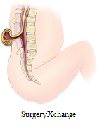
Spina bifida through SurgeryXchange
Spina bifida is a birth defect which takes place when the spine and spinal cord don't form correctly. It comes under the wide category of neural tube defects. The neural tube is the embryonic construction that ultimately develops into the baby's brain, spinal cord, and tissues that surround them. Usually, the neural tube forms early in pregnancy, and it closes by the 28th day after formation. In babies with spina bifida, a part of the neural tube fails to grow or close properly, causes the faults in the spinal cord and in the bones of the spine.
Types of Spina Bifida
  1. Meningocele
In a form of spina bifida called meningocele, the protective tissues around the spinal cord drive out through the opening in the vertebrae, creating a sac filled with liquid. But this sac doesn't contain spinal cord, so nerve injury is less likely to happen, however, later complications are possible.
  1. Spina bifida occulta
The mildest form, spina bifida occulta results in a minor separation or gap in one or more of the bones of the spine. Several people who have it don't even know it, except the condition is discovered during an imaging test done for unrelated reasons.
  1. Myelomeningocele
It is also known as open spina bifida, it is the most severe form. The spinal canal is open along with numerous vertebrae in lower or middle back. The tissues and spinal nerves push through this opening at birth, forming a sac on the baby's back, typically exposing tissues and nerves. This makes the baby prone to life-threatening infections.
Symptoms
Following are the symptoms of spina bifida:
  1. Spina bifida occulta.Since the spinal nerves generally are not involved, typically there are no signs or symptoms. However, evident signs may sometimes be seen on the newborn's skin above the spinal defect, including an abnormal tuft of hair, or a small dimple or birthmark.
  2. The membranes near the spinal cord push out through an opening in the vertebrae, creating a sac filled with fluid, but this sac does not include the spinal cord.
  3. In this severe form of spina bifida:
    1. The spinal canal remains open along several vertebrae in the lower or middle back.
    2. Both the membranes and the spinal cord or nerves protrude at birth, forming a sac.
  • Tissues and nerves usually are exposed, though sometimes skin covers the sac.
Risk and Complications
Risks include:
  • Family history of neural tube defects. Couples who have had one child with a neural tube defect have higher chances of having another baby with the same defect. The risk increases if two previous children are having the same condition.
  • Some medications. For example, anti-seizure medications, such as valproic acid (Depakene), seem to cause neural tube defects when taken during pregnancy, possibly because they interfere with the body's ability to use folate and folic acid.
  • Women having diabetes with no control over their blood sugar level have a higher risk of having a baby with spina bifida.
  • Pre-pregnancy obesity can increase the risk of neural tube birth defects, including spina bifida.

Prevention

Get folic acid first

It is important to have a good amount of folic acid in your system during the early weeks of pregnancy to prevent spina bifida. Most women don't discover that they're pregnant until this time, experts say that all women of childbearing age should take a daily supplement of 400 micrograms (mcg) of folic acid.
Various foods, including enriched bread, pasta, rice, and some breakfast cereals, are fortified with 400 mcg of folic acid per serving.

Planning pregnancy

If you are trying to conceive, most pregnancy experts believe supplementation of at least 400 mcg of folic acid a day is the best approach for women planning a pregnancy.
Your body does not absorb folate as easily as it absorbs synthetic folic acid, and most people do not get the recommended amount of folate through diet alone, so vitamin supplements are necessary to prevent spina bifida.
To eat a healthy diet, with foods rich in folate or enriched with folic acid. This vitamin is naturally in following:
  • Citrus fruits and juices.
  • Egg yolks.
  • Dark green vegetables.

Tuesday 9 October 2018

WHEN SOMETHING THAT YOU LOVE MAKES YOU WEAK IN THE KNEES!

Before your mind runs wild, let me make it clear that this isn’t completely dirty and painful. Or is it? Each person is on their own, picking up what they like and love persuading towards that particular activity. While you’re at it, doing what you love there are often times when a small misjudgment or a miscalculation might end up giving months of terrible and excruciating pain. One such incident might be A LIGAMENT TEAR.
Ligament tear usually occurs in the joints of our body when a great force is applied to a particular joint. Let’s focus on the knee joint as of now. The knee joint has 4 ligaments namely,
  • Anterior cruciate ligament(ACL)
  • Posterior cruciate ligament(PCL)
  • Medial cruciate ligament(MCL)
  • Lateral crucial ligament(LCL)
These ligaments hold the knee joint in place and help in locking and unlocking the joint. When you walk, run, dance, gym or jump these ligaments work continuously. When the knee joint is locked, the greater force applied on it ends up tearing the ligament depending upon the direction of this unaided force.
Injury to PCL – Ligament tears are also common in a road traffic accident. Usually, in a car accident, the posterior cruciate ligament is torn. The harm occurs when the tibia is forced back to the femur. This is rather rare in a sports person whose anterior cruciate ligament is at risk.
Injury to ACL – Twisting a semi-flexed knee, which already made you say “ouch” is quite cumbersome to deal with as it often causes injury to the medial meniscus and MCL. (O’Donoghue triad)
Injury to MCL – Any force the pushes the tibia towards the femur in an inside fashion causes the rupture of this ligament.
Injury to LCL – MCL, and LCL are attached to either side of the joint, media is on the inner side and lateral on the opposite. So the damage to the lateral ligament occurs when the force is applied to adduct the tibia to the femur.

So now, describing the proper anatomy. The knee joint is a hinge joint.
A hinge joint is one which is made up of more than 2 bones and moves along one axis only. Elbow joint is another such synovial joint. Knee joint consists of the tibia and the femur, a tibiofemoral joint. Then there is patella which glides over the front of the tibial end of the femur, forming a patellofemoral joint. The knee Ligaments act like the joint’s best friend, providing support and stability. When this harmony is disturbed, a painful unstable state happens to be.
The ligament injury can be associated with,
  • Chipping of the bone to which the ligament it is attached.
  • A mid-substance tear ( tear in the ligament)
  • Severity may vary on the tear being complete or incomplete.
  • One ligament involvement or more.
The combination depends upon the force acting on the joint. More severe the injury more intricate the treatment regimen gets.
Diagnosis can be made through clinical examination and a detail on the event of the grievance. Complications that present with the tear are dislocation of the joint, blood clots, swelling. The pain may be localized over the torn ligament. Radiological examination of the knee joint is important to check if the bone has been chipped off, which might have been avulsed from the ligament attachment. Contrary to the Stress X-rays, MRI is a non-invasive method to diagnose ligament injuries. The arthroscopic examination may be needed in a few cases.
Treatment of this condition was conventionally done by non-operative methods. Now that there are better techniques and with the recent advances, better results are achieved through operative reconstruction. Operative reconstruction has become popular in high demand athletic individuals like Tom Brady, Alex Morgan Adrian Peterson and a lot more NBA players. Celebrity or not we are all made up of the same cells and same anatomy and ligament injury can be treated and normal functioning of the knee can be obtained and hence decreasing your time not spent on doing what you love.
Methods that are undertaken are,
  • Conservative method – The blood clot/ hematoma is aspirated and the knee is immobilized in a cylinder cast or a knee immobilizer. If the injury is grade I or grade II, a conservative method is successful for treatment. Followed by this, strengthening physiotherapy is required.
  • Operative methods – These are to be done when there is a combination of ligament injuries, involving multiple ligaments especially in young athletes or dancers. Usually, the surgery is done 2-3 weeks after the injury when the inflammation is fairly reduced.
What do the surgeons do actually? They first go by repairing the ligament. It is performed on grade III ligament tears, which means the ligament is completely torn. If the surgery is done after the 2-3 weeks, an additional reinforcement is provided by a tendon or fascial graft as the original torn ligament would have lost the strength due to a complete tear.

Secondly, they reconstruct the unstable knee injuries which present later than usual. An allograft or an autograft of a tendon or a fascia lata is used; a synthetic graft can also be used. Auto is better than all as there are lesser chances of rejection.

Knee ligament tears are very common and are often undiagnosed. Ligament tears are usually not seen on X-rays and might get neglected. The usual presenting symptom is the instability of the knee joint. Treatment is biased depending upon the activity level of the patient. If you’re a ninja then you’ll probably need surgery. If you’re the person who probably has a sedentary lifestyle, the doctor might perhaps recommend physiotherapy. Usually, the ACL tends to get damaged more often than the others. ACL reconstruction is done with the help of an autograft and bio-absorbable screws. Arthroscopic ACL reconstruction is the surgery of choice as it is minimally invasive, and results are outstanding as the patients revert back to normal with negligible risks.
By Dr. Apoorva Manjunath.
For more information visit- https://surgeryxchange.com/

Saturday 6 October 2018

Should I undergo Knee Replacement Surgery?

Every year approximately 700,000 knee replacement procedures are performed in the United States of America. This number is projected to increase to 3.48 million procedures per year by 2030. https://www.uptodate.com/contents/total-knee-replacement-arthroplasty-beyond-the-basics) However, this number represents the tip of the iceberg. An astounding number of patients, continue to look for pain relief and non-surgical treatment options.
Most of them are advised weight loss, physiotherapy, braces, medications, and injectables. While these treatment modalities are effective at initial stages of knee arthritis, at later stages they provide limited or no relief.
Despite being in pain, patients often delay or avoid surgery. We asked our experts at SurgeryXchange, what are the most common apprehensions that patients have?
Is knee replacement surgery the right decision for me?
If your knee pain has aggravated to a point that you have pain at night or even when you’re resting, you might want to consider surgical treatment. If you have already tried medicines, injections, physiotherapy and still you’re bogged down with pain, surgery may be the only option. The most important question you need to answer is if pain or lack of mobility is hampering you from living a normal life, then yes, surgery is the answer.

What happens during knee replacement surgery?
Arthritis or osteoarthritis, which usually happens as age progresses, is a process that leads to wear and tear of the joint. The joint surface is covered by a smooth layer of a substance known as cartilage that allows smooth movement in the joint. With age, there is a breakdown of this joint cartilage. The damage, in turn, limits movement of the joint and causes pain. During knee replacement or arthroplasty, the surfaces of the knee which are damaged are removed and replaced with artificial material.

How long does it take to recover from knee replacement surgery?
The procedure last 1.5 to 2 hours. Physiotherapy is the cornerstone to recovery. Patients are discharged with 1-3 days. Patients can resume normal daily activities such as walking and bathing within several days of discharge. A trained physiotherapist assists them post-surgery and works closely with them over the next few weeks till they recover strength and mobility.

How long does the new knee last?
Scientific data suggests that for patients who got their joint replaced today have a 90-95% chance that their joint will last 10 years and an 80-85% that it will last 20 years.
(https://www.sciencedaily.com/releases/2011/02/110217082925.html)

How is life after knee replacement surgery different from the life before?
More than 90 percent of patients suffering from severe pain and inability to walk experience a dramatic reduction in knee pain and a significant improvement in the ability to perform common activities. (https://www.sciencedaily.com/releases/2011/02/110217082925.htm) But the bigger gain is the impact on quality of life. Most patients describe the surgery as transformative, as it gives excellent pain relief and provides them with the ability to be independent in their day-to-day functioning of life.
So, are you struggling with knee pain? If you’re having trouble getting up out of bed, going to the toilet or fetching yourself a glass of water, it’s time to consider surgery at www.surgeryxchange.com.  A thorough examination by an Orthopedic Surgeon should be the right next step. For booking an expert opinion, visit 
https://surgeryxchange.com/best/cost-of-total-knee-Replacement-Surgery-in-bangalore

Wednesday 3 October 2018

TYPES OF HEART SURGERY

Coronary Artery Bypass Grafting
The most common type of heart surgery is Coronary Artery Bypass Grafting (CABG). CABG improves blood flow to the heart. Surgeons use CABG to treat people who have severe coronary heart disease (CHD).
CHD is a disease in which a waxy substance called plaque builds up inside the coronary arteries. These arteries supply oxygen-rich blood to your heart.
If the plaque ruptures, a blood clot can form on its surface. A large blood clot can mostly or completely block blood flow through a coronary artery. This is the most common cause of a heart attack. A healthy artery or vein from the body is connected to the blocked coronary artery during CABG. The grafted vein or artery bypasses the blocked portion of the coronary artery. This generates a fresh path for oxygen-rich blood to flow to the heart muscle.
Surgeons can bypass multiple blocked coronary arteries during one surgery.
Transmyocardial Laser Revascularization
TMR or Transmyocardial laser revascularization is surgery used to treat angina.
In case TMR is done, the procedure may be executed through a small opening in the chest. During TMR, a surgeon uses lasers to make small channels through the heart muscle and into the heart’s lower left chamber (the left ventricle).
Heart Valve Repair or Replacement
Heart surgery is performed to repair leaflets that do not open as wide as they should. This may happen if they become stiff or thick or fuse together. As a result, not enough blood flows through the valve.
Heart surgery also is used to fix leaflets that don’t close tightly. Rather than only moving forward into the arteries as it should, this issue can make blood leak back into the heart chambers.
To fix these problems, surgeons either repair the valve or replace it with a man-made or biological valve. Biological valves are made from human, cow or pig heart tissue and may have man-made parts as well.
To repair a mitral or pulmonary valve that’s too narrow, a cardiologist will insert a catheter through a large blood vessel and guide it to the heart.
The cardiologist will put the end of the catheter in the narrow valve. They will deflate and inflate a little balloon at the tip of the catheter, to widen the valve, allowing more blood to flow through it. This approach is less invasive than open-heart surgery.
Arrhythmia Treatment
Arrhythmia is an issue with the rhythm or rate of the heartbeat.
Surgical methods are used to implant an implantable cardioverter defibrillator (ICD) or a pacemaker.
An ICD is a device which is connected to your heart with wires. After checking your heartbeat, if the device senses a dangerous arrhythmia, it sends an electric shock to your heart to restore a normal heart rhythm.
Wires connect the pacemaker to your heart chambers. The device uses low-energy electrical pulses to control your heart rhythm. Most pacemakers have a sensor that starts the device only if your heart rhythm is abnormal.
Another arrhythmia treatment is called maze surgery. For this type of surgery, the surgeon creates a fresh path for the heart’s electrical signals to travel through. This type of surgery is used to treat atrial fibrillation, the most common type of serious arrhythmia.
Aneurysm Repair
An aneurysm is a balloon-like bulge in the wall of an artery or the heart muscle. Repairing an aneurysm involves surgery to replace the weak section of the artery or heart wall with a patch or graft.
Heart Transplant
A surgery or surgical method to remove a patient’s diseased heart and swap it with a healthy heart from a deceased donor is a heart transplant. Heart failure is a condition in which the heart is damaged or weak.
Patients waiting for a donor's heart receive an ongoing treatment for heart failure and other medical conditions. Total Artificial Hearts (TAHs) or Ventricular Assist Devices (VADs) might be used to treat these patients.
Surgery to Place Total Artificial Hearts or Ventricular Assist Devices
TAH is a device that replaces the two lower chambers of the heart (the ventricles).
VAD is a mechanical pump that is used to sustain blood flow and heart function in people who have weak hearts.
Placing either device requires open-heart surgery.
Open-Heart Surgery
Open-heart surgery is a type of surgery in which a surgeon makes a large incision in the chest to open the rib cage and operate on the heart. The surgeon also may open the heart, depending on the type of surgery.
Open-heart surgery is used to place VADs and TAHs, do heart transplants, treat atrial fibrillation, replace or repair heart valves, and do CABG.
Off-Pump Heart Surgery
Surgeons also use off-pump or beating heart, surgery to do CABG. The heart isn’t stopped, and a heart-lung bypass machine isn’t used.
Minimally Invasive Heart Surgery
A surgeon makes small incisions in the side of the chest between the ribs for minimally invasive heart surgery. A heart-lung bypass machine may or may not be used in this type of surgery.
Minimally invasive heart surgery is used to do some bypass and maze surgeries. It’s also used to repair or replace heart valves, insert pacemakers or ICDs, or take a vein or artery from the body to use as a bypass graft for CABG.
Robotic-assisted surgery is a type of minimally invasive heart surgery for which a surgeon uses a computer to control surgical tools on thin robotic arms.

Tuesday 4 September 2018

What you should know about Liposuction

As celebrities such as Kerry Katona and Chrissy Teigen flaunt their newly acquired bodies, thousands of men and women around the world queue up outside the liposuction clinics, searching for the best plastic surgeons in the world.
Liposuction, also referred to as “lipo” is a type of cosmetic surgery that reshapes specific areas of the body by removing excess fat deposits and improving your body contours.
Before you succumb to this fad, you should know a few facts about Liposuction:
1. You can get Liposuction anywhere on your body
Though traditionally performed on the hips, belly, thighs, buttocks, it can be done anywhere on your body. The most frequently treated areas for women are the abdomen, breasts, hips, outer thighs, front of thighs, inside of thighs, around the knees, upper arms, buttocks, cheeks, armpits, and neck. Men comprise about 15% to 25% of liposuction candidates, and frequently seek a reduction of fat in the chin and neck area, abdomen, flanks, and breasts. Liposuction cost in India is affordable.
2. Yes, Liposuction permanently removes fat
During Liposuction, fat cells are removed permanently. While fat regrowth does not usually occur in the treated area, the body constantly redistributes fat and hence, if the individual does not follow a healthy exercise and diet regimen, he or she may regain the weight or lose the contours.
3. Recovery is Gradual
Liposuction often leaves swelling and inflammation in the area treated, and it could be a couple of months before you have the desired results. While you can return to day to day activities immediately, the bruising, swelling and soreness take a few weeks to subside.
4. Plastic Surgeons and Dermatologists Perform Most Liposuctions
The web is full of doctors and beauty centers offer liposuction services. It is pertinent to ask for training certifications and scientific affiliations before finalizing a provider. Don’t be shy about getting a second option or asking the right questions about the process and expertise.
5. Liposuction is not for the overweight or obese
Liposuction is not an alternative to losing weight. Instead, it should be considered as re-shaping surgery and should be used to reduce spot or stubborn fat, where diet and exercise do not reach. In fact, large volume liposuction can lead to an increase in the risk of serious surgical complications. Moreover, studies have linked the removal of more than 11 pounds or 5 Litres of fat with fluid imbalance and death.
6. Make an informed decision
Once you’ve decided on getting a liposuction, the next important decision to make is where to get it done. Untrained hands and improper removal may cause the skin to appear bumpy or withered. This is, unfortunately, the most common complication of the liposuction procedure, and sadly it is also permanent. Less common complications include fluid accumulation under the skin, temporary or permanent numbness, skin infection. One of the rare but fatal complications of Liposuction is fat embolism, where a piece of loosened fat might travel to the lungs or brain, resulting in fatal consequences.

Monday 3 September 2018

Cosmetic surgery

Cosmetic surgery or Aesthetic surgery is a unique surgical procedure that focuses on enhancing the appearance through corrections by surgical and medical techniques. Cosmetic surgery can be performed on all parts of the body. Under cosmetic surgery, there are various surgeries and reconstruction surgery is a part of cosmetic surgery. Its focus is to improve function and to give a normal appearance to a part of the person’s body.
Rhinoplasty
This is referred to a surgical treatment that reshapes or resizes the nose. It can correct a variety of concerns, including a noticeable bump on the nasal bridge, a droopy or expanded nasal tip, a nose that’s off-center, or lack of balance due to a previous injury. After the evaluation, he or she can tell the doctor if the assumptions are realistic. The surgeon will make changes based on the overall health of the person.
While initial swelling would last for a few weeks, it may take up to a year for the new nasal contour to fully refine. During this time one may notice gradual changes in the appearance of the nose as it refines to a more permanent outcome.
As the body ages, it is natural to have some changes to your face progressively, including your nose. But most of the time it will be a permanent result. A healthy lifestyle and life-long sun protection will help extend the results of the corrected appearance.
Abdominoplasty
Deep underneath the skin lies a layer of muscle that can become stretched out with age, pregnancy and weight alteration. No matter how much exercise one does, if the underlying tissues are not in good position, the stomach does not appear to be thin, trim and contoured.
Even if there is little fat on the stomach, if the muscles have detached, the lower section of the tummy will most likely look bloated and barrel-shaped. That’s because the muscles that provide the stomach definition—the six-pack muscles—have detached internally. Most surgeons say the main reason for this in women is because of pregnancy. Once the muscles are separated, they create this minor bulge. Post-pregnancy, the muscles can get stronger, but they can’t settle back to their original shape and position. The only way to correct the problem is to surgically tighten the muscles, with a tummy tuck, which in turn will remove the pooch.
Liposuction
Removes fat from your body using suction. During liposuction, small, thin, blunt-tipped tubes are inserted through minor cuts in the skin. Fat is siphoned out through these tubes as the surgeon moves the tube around and under the skin to target specific fat deposits.
In recent years, improved procedures have made liposuction safer, easier, and less painful.
Liposuction treatment has both advantages and disadvantages. It depends on how a patient recovers or how his metabolism responds to recover. Removal of excess fat can be harmful to the patient must choose the right hospital and see how well the doctor is qualified to treat him or her.
Post Liposuction
After the procedure, the area of the body that was treated is nicely wrapped to help reduce swelling, bruising, and pain. They may have to wear the compression cloth or wrap it around the area of surgery for 3 to 4 weeks.
Why Liposuction is done
The main reason for liposuction is to reshape one or more areas of the body, not to reduce body mass. Liposuction is typically used on “problem” areas that have not responded well to diet & exercise. These areas are often on the outer thighs and hips on women and the waist and back on men. The face, neck, abdomen, back, buttocks, legs, and upper arms are all commonly treated areas.
Liposuction is sometimes used in combination with other cosmetic surgery procedures, such as like tummy tuck and breast reduction treatments
Liposuction may also be used to treat certain medical conditions, including:
Lipoma is a benign tumor which is composed, basically, it is a body fat
Abnormal growth of male breast
Problems of metabolism
Armpit Sweating
Liposuction is not used to treat obesity. It will not eliminate cellulite or stretch marks.
Circumferential Thighs or Buttocks
Liposuction circumferential thighs and buttocks are referred to the reshaping of the entire thigh and buttocks region area by eliminating excess fat deposits. This technique is a cosmetic surgery procedure designed to enhance the lower body appearance and is common for individuals who have recently lost an excessive amount of weight, for those who wish to remove excess fat, for those that cannot lose the last amount of weight in their lower bodies on their own and following childbirth. Liposuction is a common technique that can be used for almost any area of the body. It is not a procedure that is designed to help someone to lose weight or to replace healthy behaviors.
The first step in a circumferential thigh and buttocks liposuction procedure is to control anesthesia and this will be evaluated through the anesthesiologist. The amount of anesthesia to be given will be determined by the estimated time of surgery, the height and weight of the patient and their overall health status.
Then, the incisions will be made. Liposuction is performed using tiny incisions, often in multiple locations along the side of the thighs and underneath the buttocks in the natural crease area. The surgeon will work according to the plan to limit the number of incisions while still being effective in the procedure.
The sterile solution is first injected into the incisions as a way to reduce the overall bleeding during the surgery as this will ultimately reduce trauma. A hollow tube is then inserted into the incision sites. A forward and backward motion is used by the surgeon to loosen the fat so that it can be sucked out using a vacuum-like machine. Having done with thighs and buttocks liposuction, the movements are often much bigger than a more gentle procedure such as facial liposuction. Many times as the incisions are so small, they can be closed with surgical tape or surgical adhesive. Bandages are often tightly wrapped around the areas affected, to help reduce swelling.
Breast Reduction
Sometimes liposuction is used along with surgery. If most of the breast is thick tissue and if excess skin isn’t a problem, liposuction alone may be enough for breast reduction.
Breast reduction surgery is done in a hospital or surgical center, typically with general anesthesia. The surgery is carried out in about 3 to 5 hours. An overnight stay is not usually required at the hospital. For minor reductions, the surgery may be done with local anesthesia.
Breast lift (mastopexy)
Same as breast reduction, except that in some cases only skin is taken out. A breast lift can raise sagging or drooping breasts, which is a common problem with large, heavy breasts, and can elevate the nipple and areola.
What To Expect After Surgery
Soon after surgery, a thin transparent fabric of silk is placed above the minor slit, and the breasts are wrapped in an elastic bandage. In some cases, there may be a tiny tube in each breast to allow blood to flow outside and fluid for the first two days. Stitches may be eliminated in 1 to 2 weeks.
Nearly all women have some breast discomfort for the first couple of days after surgery and then little discomfort for a week or two. Medicine can help soothe the pain. Swelling and bruising will be there for several weeks. Wearing a surgical bra for the whole day can help reduce swelling and support the breasts while they heal.
One can be back to normal work and social activities within a couple of weeks unless those activities involve heavy lifting or movements for the breast. The treated person may need to take a break from exercise and activities for weeks or more. It’s important to wear a bra that supports the breasts well, such as a sports bra.
You will have noticeable scars on your breasts after breast reduction surgery. Scars may fade over time, but they will not disappear.
Blepharoplasty
Eyelid surgery, or blepharoplasty, is a surgical procedure to improve the appearance of the eyelids. Surgery can be performed on the upper lids, lower lids or both. If one wants to improve the appearance or are experiencing functional problems with the eyelids, with an eyelid surgery it can rejuvenate the area surrounding your eyes.
What eyelid surgery can treat
Loose or sagging skin that creates folds or disturbs the natural contour of the upper eyelid, sometimes impairing vision
Fatty deposits that appear as puffiness in the eyelids
Bags under the eyes
Drooping lower eyelids that reveal right below the iris
Excess skin and fine wrinkles of the lower eyelid

Friday 31 August 2018

How is Rhinoplasty safe in India?

Rhinoplasty is the most complicated and challenging procedure in all of the plastic surgery. the nose job should sync to the look of the face, but also each area of the nose must be harmonious with all the others. For example, eliminating a bump without changing the tip in most cases can make the tip appear like it’s sticking out too far from the face. Contrarily, changing the tip alone without making the bridge can make a bridge appear too high or too wide. A rhinoplasty physician will go through carefully before performing the procedure.
Certainly, poorly performed work can look un-natural or look like it is artificially done. A normal example of this is the overly strained and upturned tip. In years past, the cartilage of the nose was frequently over-reduced or eliminated which can result not only in this appearance but also in deprived breathing. A better rhinoplasty, on the other hand, typically looks natural, is no longer a distracting feature, and draws people’s attention to the person’s eyes, smile, hair, and skin. An expected rhinoplasty patient should have original expectations. Each person’s face and appearance is different. Here is the list of other cosmetic procedures Brad Pitt’s nose would not appear good on every man face is it achievable. Blake Lively’s nose would not be great for every woman.
Would you want a general constructor doing complex plumbing or electrical work? Do you really want someone who does pre-supreme breast augmentations and tummy tucks executing your rhinoplasty? Rhinoplasty is an exceedingly complicated and demanding operation. A millimeter or two can make a major difference in the result. It should be performed by someone who is skilled and has specific training in it. Typically, the physician should be board certified in facial plastic and reconstructive surgery (and have a specific interest in performing rhinoplasty). This physician will have more concern about the beauty of the nose and face, but also he or she should have a great knowledge and understanding of the many functional parts of this complex organ. (e.g. breathing, sense of smell, mucus or sinus issues).

Tuesday 21 August 2018

Keep your bones healthy with the help of nutrition foods

Bone is the core important part of your body it is a structure that supports and keeps your body parts composed like an automobiles chassis which keeps the engine and other important parts together. to keep up with healthy bones there are some nutrition methods which are listed in the later part of the blog. Your body is developed based on your bones growth and nutrition for each and every physical activity you do the bones participation is 100%, to maintain a healthy life the bones play important roles.
In childhood, most of our bone growth and bone density happens. Girls reach their maximum bone density by age 18. For boys, this happens by age 20. In adulthood, it is important to maintain bone density and try to slow the rate of bone loss. As we grow older, our bodies lose bone density.

To have a healthy bone you should keep your diet plan like this.

Keep a diet plan that you can consume enough Potassium, vitamin K and magnesium which helps your body to absorb and use calcium. keeping your bone healthy results is high stamina and you can look fit. Get these important nutrients by eating a variety of healthy foods like vegetables and fruit, legumes (beans, peas, lentils), nuts, seeds, whole grains, and fish. Protein helps to build muscle, which helps keep bones strong. Choose protein-rich foods such as fish, shellfish, legumes (beans, peas, lentils), nuts, seeds and grains like wheat, horse-grams with mung beans (green grams).

Include these lists in your diet which are mainly seafood and dairy products

Milk, Fortified soy and rice beverages, Fortified orange juice, Fatty fish like salmon and sardines, Margarine, Egg yolks etc.
Include physical activity in your daily routine. Weight-bearing exercises, such as walking, jogging, tennis and climbing stairs, can help you build strong bones and slow bone loss. To have a life without physical activity it is like piling up your fat and stuffing up your muscles with unwanted fats which result is bone fractures or it may lose your bones density keep your bones healthy because it is a core part of your body you can read other blogs https://surgeryxchange.com/blog

Saturday 18 August 2018

Gastroenterology

The core objective of medical and surgical gastroenterology is focused on, diagnosis, treatment, and prevention of diseases pertaining to the liver and pancreatico-biliary system, digestive tract, in both children and adults.SurgeryXchange helps users to discover state-of-the-art traditional and endoscopic surgical procedures for gastrointestinal bleeding, gastrointestinal cancers, and foreign body removal. Our list of hospitals has  top specialist surgeons who  manage various types of  gastrointestinal surgical problems relating to the hepatobiliary tract, intestines, pancreas, and liver through  technologically advanced procedures and also offered at low-cost surgeries in India
What is Gastroenterology?
Gastroenterology is a branch of medicine dedicated to the study, diagnosis, and treatment of the gastrointestinal tract and the accessory organs of digestion. It comprises the oral cavity, the esophagus, the stomach, the small intestine, the large intestine, and the rectum. The accessory organs of digestion are pancreas, the liver, and gallbladder. Gastroscopy cost in India through SurgeryXchange is just a fraction when compared to cost in other developed countries. Additionally, medical facilities in India match to that of international standards and are renowned globally to being one of the best places and also offering low cost of surgeries in  India.
Cost of Gastrointestinal Surgeries in India
India is the most sought out destination for Gastrointestinal Surgeries. The Gastrointestinal Surgeries costs in India are affordable and just a fraction of what you normally pay in the western developed countries. Offering high-tech and advanced medical solutions to a large variety of health care problems, it is no surprise that India is one of the most preferred global healthcare destinations and SurgeryXchange is your preferred partner for your medical tour.
Cholecystectomy is Removal of a diseased gallbladder. Gallstones are also a major cause of worry, especially among obese patients. Generally, gallstone formation is influenced by a number of factors, also mandates for surgery. Gall Bladder surgery in India is affordable and SurgeryXchange helps you find best in treatment and relatively low cost than in the developed world.
Low-Cost Gastric Surgeries in India
Anal Fissures procedure
Rectal or Anal or fissure is normally a break or tears in anal canal skin. These fissures can be noticed causing bleeding on toilet paper or bright redness of anus. In acute or severe cases these cause severe pain post-defecation. Fissure depths can in cases extend to underlying sphincter muscle or be superficial in cases. If medical therapy has failed to heal for 3 months or more then surgical procedures are applied for the treatment of anal fissures. Generally, this surgery for anal fissures is done under anesthesia in cases where the anal sphincter muscle is most likely to get incised. This surgery could be painful post-surgery until the time of complete recovery. India offers a plethora of options to get low-cost anal fissures procedure. SurgeryXchange listed hospitals offer the procedure and surgeries in India that are recognized best in service and standards.
Appendectomy
Appendicectomy or Appendectomy is primarily the surgical removal of the vermiform appendix. Generally, this surgery is performed in emergencies where patients are suffering from acute conditions of appendicitis, in case of doubt one can apply minimally invasive laparoscopic procedures that are useful for diagnosis. Even though it is expensive and resource intensive compared to open surgery, recovery is quicker with such laparoscopy procedures.T here are various hospitals that you can find on SurgeryXchange to get the best options and low-cost surgeries and medical tour.
Colectomy
Colectomy is also an occasional term used to describe entire large intestine removal along with rectum (proctocolectomy) which consists of surgical resection of colon. Some of the common indications for Colostomy generally include diverticular disease of large intestine and diverticulitis, trauma, colon cancer typhlitis, bowel infarction and inflammatory bowel diseases like ulcerative colitis and Crohn’s disease. It’s our endeavour at SurgeryXchange to offer the best options for surgeries in India that are low cost and best in service.

ಸೀಳು ತುಟಿಯ ಬಗ್ಗೆ ಕೇಳಿದ್ದೀರಾ?

ಸೀಳು ತುಟಿ  ಮೇಲಿನ ತುಟಿಯ ಭಾಗದಲ್ಲಿ ಸೀಳಿಕೊಂಡಿದ್ದರೆ ಅದನ್ನು ಸೀಳು ತುಟಿ ಮತ್ತು ಸೀಳು ಅಂಗುಳ  ಎಂದು ಕರೆಯಬಹುದು.  ಮುಖದ ರಚನೆಯು ಸರಿಯಾಗಿ ಬೆಳವಣಿಗೆಯಾಗದಿದ್ದರೆ ...